Our brains take rhythmic snapshots of the world as we walk – and we never knew

For decades, psychology departments around the world have studied human behaviour in darkened laboratories that restrict natural movement.

Our new study, published today in Nature Communications, challenges the wisdom of this approach. With the help of virtual reality (VR), we have revealed previously hidden aspects of perception that happen during a simple everyday action – walking.

We found the rhythmic movement of walking changes how sensitive we are to the surrounding environment. With every step we take, our perception cycles through “good” and “bad” phases.

This means your smooth, continuous experience of an afternoon stroll is deceptive. Instead, it’s as if your brain takes rhythmic snapshots of the world – and they are synchronised with the rhythm of your footfall.

The next step in studies of human perception

In psychology, the study of visual perception refers to how our brains use information from our eyes to create our experience of the world.

Typical psychology experiments that investigate visual perception involve darkened laboratory rooms where participants are asked to sit motionless in front of a computer screen.

Often, their heads will be fixed in position with a chin rest, and they will be asked to respond to any changes they might see on the screen.

This approach has been invaluable in building our knowledge of human perception, and the foundations of how our brains make sense of the world. But these scenarios are a far cry from how we experience the world every day.

This means we might not be able to generalise the results we discover in these highly restricted settings to the real world. It would be a bit like trying to understand fish behaviour, but only by studying fish in an aquarium.

Instead, we went out on a limb. Motivated by the fact our brains have evolved to support action, we set out to test vision during walking – one of our most frequent and everyday behaviours.

Doing tests in a lab isn’t quite the same as seeing and interacting with things in the real world. sirtravelalot/Shutterstock

A walk in a (virtual) forest

Our key innovation was to use a wireless VR environment to test vision continuously while walking.

Several previous studies have examined the effects of light exercise on perception, but used treadmills or exercise bikes. While these methods are better than sitting still, they don’t match the ways we naturally move through the world.

Instead, we simulated an open forest. Our participants were free to roam, yet unknown to them, we were carefully tracking their head movement with every step they took.

Participants walked in a virtual forest while trying to detect brief visual ‘flashes’ in the moving white circle.

We tracked head movement because as you walk, your head bobs up and down. Your head is lowest when both feet are on the ground and highest when swinging your leg in-between steps. We used these changes in head height to mark the phases of each participant’s “step-cycle”.

Participants also completed our visual task while they walked, which required looking for brief visual “flashes” they needed to detect as quickly as possible.

By aligning performance on our visual task to the phases of the step-cycle, we found visual perception was not consistent.

Instead, it oscillated like the ripples of a pond, cycling through good and bad periods with every step. We found that depending on the phases of their step-cycle, participants were more likely to sense changes in their environment, had faster reaction times, and were more likely to make decisions.

Oscillations in nature, oscillations in vision

Oscillations in vision have been shown before, but this is the first time they have been linked to walking.

Our key new finding is these oscillations slowed or increased to match the rhythm of a person’s step-cycle. On average, perception was best when swinging between steps, but the timing of these rhythms varied between participants. This new link between the body and mind offers clues as to how our brains coordinate perception and action during everyday behaviour.

Next, we want to investigate how these rhythms impact different populations. For example, certain psychiatric disorders can lead to people having abnormalities in their gait.

There are further questions we want to answer: are slips and falls more common for those with stronger oscillations in vision? Do similar oscillations occur for our perception of sound? What is the optimal timing for presenting information and responding to it when a person is moving?

Our findings also hint at broader questions about the nature of perception itself. How does the brain stitch together these rhythms in perception to give us our seamless experience of an evening stroll?

These questions were once the domain of philosophers, but we may be able to answer them, as we combine technology with action to better understand natural behaviour.The Conversation

Matthew Davidson, Postdoctoral research fellow, lecturer, University of Sydney

This article is republished from The Conversation under a Creative Commons license. Read the original article.

Read More........

How exercise increases brain volume — and may slow memory decline

Exercising for 25 minutes a week, or less than four minutes a day, could help to bulk up our brains and improve our ability to think as we grow older. A new study, which involved scanning the brains of more than 10,000 healthy men and women from ages 18 to 97, found that those who walked, swam, cycled or otherwise worked out moderately for 25 minutes a week had bigger brains than those who didn’t, whatever their ages.

Bigger brains typically mean healthier brains.

The differences were most pronounced in parts of the brain involved with thinking and memory, which often shrink as we age, contributing to risks for cognitive decline and dementia.

“This is an exciting finding and gives us more fuel for the idea that being physically active can help maintain brain volume across the life span,” said David Raichlen, a professor of biological sciences and anthropology at the University of Southern California. He studies brain health but was not involved with the new study.

The results have practical implications, too, about which types of exercise seem best for our brain health and how little of that exercise we may really need.

– – –

Little exercise, big brain

“We wondered, if we chose a very low threshold of exercise what would we see?” said Cyrus A. Raji, an associate professor of radiology and neurology at Washington University in St. Louis, who led the new study.

He and his colleagues were well aware that exercise is good for brains, especially as we age. Physically active older people are far less likely than the sedentary to develop Alzheimer’s disease or other types of memory loss and cognitive decline.

But he also knew that few people in the real world exercise much. “You hear that you need 10,000 steps a day,” he said, “or 150 minutes a week. But it’s very hard to reach” those goals.

Would less – even far less – exercise still help to build healthier brains, he and his colleagues wondered?

What about, for instance, 25 minutes of exercise a week, a sixth of the 150 minutes recommended in most formal exercise guidelines?

“It seemed an achievable amount for most people,” Raji said. But would it show effects on brains?

– – –

10,125 brain scans

He and his colleagues turned to existing brain scans for 10,125 mostly healthy adults of all ages who’d come to the university medical center for diagnostic tests. Beforehand, these patients had provided information about their medical histories and how often and strenuously they’d exercised during the past two weeks.

The researchers divided them into those who’d exercised for at least 25 minutes a week and those who hadn’t.

Then, with the aid of artificial intelligence, they began comparing scans and exercise habits, looking for differences in brain volume, or how much space a brain and its constituent parts fill. More volume is generally desirable.

A clear pattern quickly emerged. Men and women, of any age, who exercised for at least 25 minutes a week showed mostly greater brain volume than those who didn’t. The differences weren’t huge but were significant, Raji said, especially when the researchers looked deeper inside the organ.

There, they found that exercisers possessed greater volume in every type of brain tissue, including grey matter, made up of neurons, and white matter, the brain’s wiring infrastructure, which supports and connects the thinking cells.

More granularly, the exercisers tended to have a larger hippocampus, a portion of the brain essential for memory and thinking. It usually shrinks and shrivels as we age, affecting our ability to reason and recall.

They also showed larger frontal, parietal and occipital lobes, which, together, signal a healthy, robust brain.

– – –

Moderate exercise was best for brains

“It was surprising and encouraging” to see such widespread effects in the brains of people who were exercising so little, Raji said.

Of course, this study was associational, meaning it showed links between exercise and brain health, but not that exercise necessarily caused the improvements. So it’s possible other lifestyle factors or genetics were at play, or that people with big brains just happened to like exercise. But given the number of scans and the wide age range, Raji believes the effects of exercise on people’s brains were real and direct and would help to maintain our ability to think well as we grow older.

Exactly how exercise might be altering brains is impossible to say from this study. But Raji and his colleagues believe exercise reduces inflammation in the brain and also encourages the release of various neurochemicals that promote the creation of new brain cells and blood vessels.

In effect, exercise seems to help build and bank a “structural brain reserve,” he said, a buffer of extra cells and matter that could protect us somewhat from the otherwise inevitable decline in brain size and function that occurs as we age. Our brains may still shrink and sputter over the years. But, if we exercise, this slow fall starts from a higher baseline.

Perhaps best of all, the most effective exercise in the study was also relatively gentle. People who said they exercised moderately, meaning they could still chat as they worked out, wound up with somewhat greater brain volume than those who exercised more vigorously, such as by swift running.

But the numbers of vigorous exercisers were quite small, making comparisons suspect, Raji said, and their brain volume was still larger than among those who rarely, if ever, exercised at all.

Overall, any exercise of any type and in even small amounts is likely to be “a very good idea” for brain health, he said.Raichlen agrees. “Studies like this continue to provide strong evidence that moving your body even a small amount may have an impact on brain health, and that it is never too early, or too late, to start.”How exercise increases brain volume — and may slow memory declineImage Link Flickr
Read More........

Think you’re good at multi-tasking? Here’s how your brain compensates – and how this changes with age

Arlington Research/Unsplash Peter Wilson, Australian Catholic UniversityWe’re all time-poor, so multi-tasking is seen as a necessity of modern living. We answer work emails while watching TV, make shopping lists in meetings and listen to podcasts when doing the dishes. We attempt to split our attention countless times a day when juggling both mundane and important tasks.

But doing two things at the same time isn’t always as productive or safe as focusing on one thing at a time.

The dilemma with multi-tasking is that when tasks become complex or energy-demanding, like driving a car while talking on the phone, our performance often drops on one or both.

Here’s why – and how our ability to multi-task changes as we age.

Doing more things, but less effectively

The issue with multi-tasking at a brain level, is that two tasks performed at the same time often compete for common neural pathways – like two intersecting streams of traffic on a road.

In particular, the brain’s planning centres in the frontal cortex (and connections to parieto-cerebellar system, among others) are needed for both motor and cognitive tasks. The more tasks rely on the same sensory system, like vision, the greater the interference.

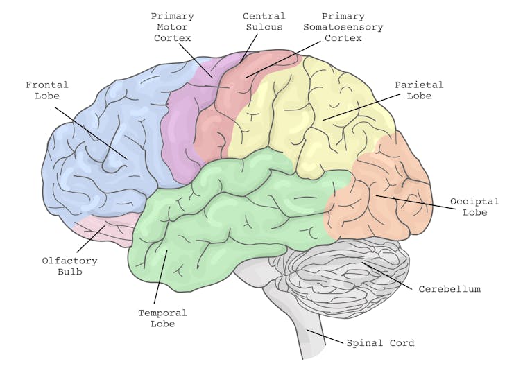
The brain’s action planning centres are in the frontal cortex (blue), with reciprocal connections to parietal cortex (yellow) and the cerebellum (grey), among others. grayjay/Shutterstock

This is why multi-tasking, such as talking on the phone, while driving can be risky. It takes longer to react to critical events, such as a car braking suddenly, and you have a higher risk of missing critical signals, such as a red light.

The more involved the phone conversation, the higher the accident risk, even when talking “hands-free”.

Having a conversation while driving slows your reaction time. GBJSTOCK/Shutterstock

Generally, the more skilled you are on a primary motor task, the better able you are to juggle another task at the same time. Skilled surgeons, for example, can multitask more effectively than residents, which is reassuring in a busy operating suite.

Highly automated skills and efficient brain processes mean greater flexibility when multi-tasking.

Adults are better at multi-tasking than kids

Both brain capacity and experience endow adults with a greater capacity for multi-tasking compared with children.

You may have noticed that when you start thinking about a problem, you walk more slowly, and sometimes to a standstill if deep in thought. The ability to walk and think at the same time gets better over childhood and adolescence, as do other types of multi-tasking.

When children do these two things at once, their walking speed and smoothness both wane, particularly when also doing a memory task (like recalling a sequence of numbers), verbal fluency task (like naming animals) or a fine-motor task (like buttoning up a shirt). Alternately, outside the lab, the cognitive task might fall by wayside as the motor goal takes precedence.

Brain maturation has a lot to do with these age differences. A larger prefrontal cortex helps share cognitive resources between tasks, thereby reducing the costs. This means better capacity to maintain performance at or near single-task levels.

The white matter tract that connects our two hemispheres (the corpus callosum) also takes a long time to fully mature, placing limits on how well children can walk around and do manual tasks (like texting on a phone) together.

For a child or adult with motor skill difficulties, or developmental coordination disorder, multi-tastking errors are more common. Simply standing still while solving a visual task (like judging which of two lines is longer) is hard. When walking, it takes much longer to complete a path if it also involves cognitive effort along the way. So you can imagine how difficult walking to school could be.

What about as we approach older age?

Older adults are more prone to multi-tasking errors. When walking, for example, adding another task generally means older adults walk much slower and with less fluid movement than younger adults.

These age differences are even more pronounced when obstacles must be avoided or the path is winding or uneven.

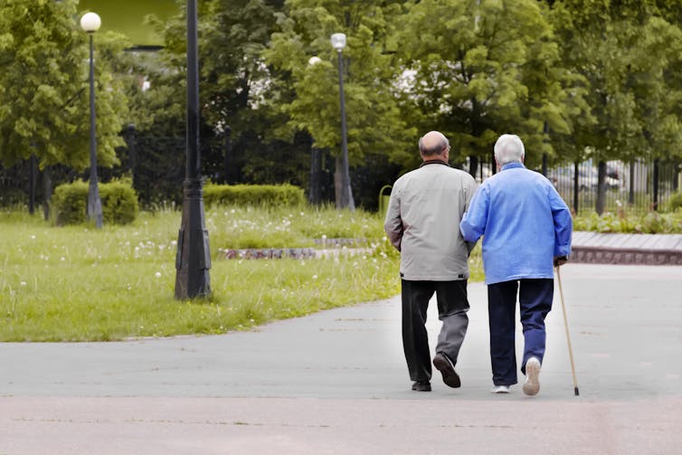
Our ability to multi-task reduces with age. Shutterstock/Grizanda

Older adults tend to enlist more of their prefrontal cortex when walking and, especially, when multi-tasking. This creates more interference when the same brain networks are also enlisted to perform a cognitive task.

These age differences in performance of multi-tasking might be more “compensatory” than anything else, allowing older adults more time and safety when negotiating events around them.

Older people can practise and improve

Testing multi-tasking capabilities can tell clinicians about an older patient’s risk of future falls better than an assessment of walking alone, even for healthy people living in the community.

Testing can be as simple as asking someone to walk a path while either mentally subtracting by sevens, carrying a cup and saucer, or balancing a ball on a tray.

Patients can then practise and improve these abilities by, for example, pedalling an exercise bike or walking on a treadmill while composing a poem, making a shopping list, or playing a word game.

The goal is for patients to be able to divide their attention more efficiently across two tasks and to ignore distractions, improving speed and balance.

There are times when we do think better when moving

Let’s not forget that a good walk can help unclutter our mind and promote creative thought. And, some research shows walking can improve our ability to search and respond to visual events in the environment.

But often, it’s better to focus on one thing at a time

We often overlook the emotional and energy costs of multi-tasking when time-pressured. In many areas of life – home, work and school – we think it will save us time and energy. But the reality can be different.

Multi-tasking can sometimes sap our reserves and create stress, raising our cortisol levels, especially when we’re time-pressured. If such performance is sustained over long periods, it can leave you feeling fatigued or just plain empty.

Deep thinking is energy demanding by itself and so caution is sometimes warranted when acting at the same time – such as being immersed in deep thought while crossing a busy road, descending steep stairs, using power tools, or climbing a ladder.

So, pick a good time to ask someone a vexed question – perhaps not while they’re cutting vegetables with a sharp knife. Sometimes, it’s better to focus on one thing at a time.The Conversation

Peter Wilson, Professor of Developmental Psychology, Australian Catholic University

This article is republished from The Conversation under a Creative Commons license. Read the original article.

Read More........

Pigs with human brain cells and biological chips: how lab-grown hybrid lifeforms bamboozle scientific ethics


Earlier this month, scientists at the Guangzhou Institutes of Biomedicine and Health announced they had successfully grown “humanised” kidneys inside pig embryos.

The scientists genetically altered the embryos to remove their ability to grow a kidney, then injected them with human stem cells. The embryos were then implanted into a sow and allowed to develop for up to 28 days.

The resulting embryos were made up mostly of pig cells (although some human cells were found throughout their bodies, including in the brain). However, the embryonic kidneys were largely human.

This breakthrough suggests it may soon be possible to generate human organs inside part-human “chimeric” animals. Such animals could be used for medical research or to grow organs for transplant, which could save many human lives.

But the research is ethically fraught. We might want to do things to these creatures we would never do to a human, like kill them for body parts. The problem is, these chimeric pigs aren’t just pigs – they are also partly human.

If a human–pig chimera were brought to term, should we treat it like a pig, like a human, or like something else altogether?

Maybe this question seems too easy. But what about the idea of creating monkeys with humanised brains?

Chimeras are only one challenge among many

Other areas of stem cell science raise similarly difficult questions.

In June, scientists created “synthetic embryos” – lab-grown embryo models that closely resemble normal human embryos. Despite the similarities, they fell outside the scope of legal definitions of a human embryo in the United Kingdom (where the study took place).

Like human–pig chimeras, synthetic embryos straddle two distinct categories: in this case, stem cell model and human embryo. It is not obvious how they should be treated.

In the past decade, we have also seen the development of increasingly sophisticated human cerebral organoids (or “lab-grown mini-brains”).

Unlike synthetic embryos, cerebral organoids don’t mimic the development of a whole person. But they do mimic the development of the part that stores our memories, thinks our thoughts, and makes conscious experience possible.

A network of neural cells grown on an array of electrodes to produce a ‘biological computer chip’. Cortical Labs

Most scientists think current “mini-brains” are not conscious, but the field is developing rapidly. It is not far-fetched to think a cerebral organoid will one day “wake up”.

Complicating the picture even further are entities that combine human neurons with technology – like DishBrain, a biological computer chip made by Cortical Labs in Melbourne.

How should we treat these in vitro brains? Like any other human tissue culture, or like a human person? Or perhaps something in between, like a research animal?

A new moral framework

It might be tempting to think we should settle these questions by slotting these entities into one category or another: human or animal, embryo or model, human person or mere human tissue.

This approach would be a mistake. The confusion sparked by chimeras, embryo models, and in vitro brains shows these underlying categories no longer make sense.

We are creating entities that are neither one thing nor the other. We cannot solve the problem by pretending otherwise.

We would also need good reasons to classify an entity one way or another.

Should we count the proportion of human cells to determine whether a chimera counts as an animal or a human? Or should it matter where the cells are located? What matters more, brain or buttocks? And how can we work this out?

Moral status

Philosophers would say these are questions about “moral status”, and they have spent decades deliberating on what kinds of creatures we have moral duties to, and how strong these duties are. Their work can help us here.

For example, utilitarian philosophers see moral status as a matter of whether a creature has any interests (in which case it has moral status), and how strong those interests are (stronger interests matter more than weaker ones).

On this view, so long as an embryo model or brain organoid lacks consciousness, it will lack moral status. But if it develops interests, we need to take these into account.

Similarly, if a chimeric animal develops new cognitive abilities, we need to reconsider our treatment of it. If a neurological chimera comes to care about its life as much as a typical human does, then we should hesitate to kill it just as much as we would hesitate to kill a human.

This is just the beginning of a bigger discussion. There are other accounts of moral status, and other ways of applying them to the entities stem cell scientists are creating.

But thinking about moral status sets us down the right path. It fixes our minds on what is ethically significant, and can begin a conversation we badly need to have.The Conversation

Julian Koplin, Lecturer in Bioethics, Monash University & Honorary fellow, Melbourne Law School, Monash University

This article is republished from The Conversation under a Creative Commons license. Read the original article.

Read More........

How consciousness may rely on brain cells acting collectively – new psychedelics research on rats

Psychedelics can help uncover consciousness. agsandrew/Shutterstock Pär Halje, Lund University
Psychedelics are known for inducing altered states of consciousness in humans by fundamentally changing our normal pattern of sensory perception, thought and emotion. Research into the therapeutic potential of psychedelics has increased significantly in the last decade. While this research is important, I have always been more intrigued by the idea that psychedelics can be used as a tool to study the neural basis of human consciousness in laboratory animals. We ultimately share the same basic neural hardware with other mammals, and possibly some basic aspects of consciousness, too. So by examining what happens in the brain when there’s a psychedelically induced change in conscious experience, we can perhaps glean insights into what consciousness is in the first place.We still don’t know a lot about how the networks of cells in the brain enable conscious experience. The dominating view is that consciousness somehow emerges as a collective phenomenon when the dispersed information processing of individual neurons (brain cells) is integrated as the cells interact.But the mechanism by which this is supposed to happen remains unclear. Now our study on rats, published in Communications Biology, suggests that psychedelics radically change the way that neurons interact and behave collectively.Our study compared two different classes of psychedelics in rats: the classic LSD type and the less-typical ketamine type (ketamine is an anaesthetic in larger doses). Both classes are known to induce psychedelic experiences in humans, despite acting on different receptors in the brain. Exploring brain waves: We used electrodes to simultaneously measure electrical activity from 128 separate areas of the brain of nine awake rats while they were given psychedelics. The electrodes could pick up two kinds of signals: electrical brain waves caused by the cumulative activity in thousands of neurons, and smaller transient electrical pulses, called action potentials, from individual neurons. The classic psychedelics, such as LSD and psilocybin (the active ingredient in magic mushrooms), activates a receptor in the brain (5-HT2A) which normally binds to serotonin, a neurotransmitter that regulates mood and many other things. Ketamine, on the other hand, works by inhibiting another receptor (NMDA), which normally is activated by glutamate, the primary neurotransmitter in the brain for making neurons fire. We speculated that, despite these differences, the two classes of psychedelics might have similar effects on the activity of brain cells. Indeed, it turned out that both drug classes induced a very similar and distinctive pattern of brain waves in multiple brain regions. The brain waves were unusually fast, oscillating about 150 times per second. They were also surprisingly synchronised between different brain regions. Short bursts of oscillations at a similar frequency are known to occur occasionally under normal conditions in some brain
Brain waves on electroencephalogram EEG. Chaikom/Shutterstock
regions. But in this case, it occurred for prolonged durations.  First, we assumed that a single brain structure was generating the wave and that it then spread to other locations. But the data was not consistent with that scenario. Instead, we saw that the waves went up and down almost simultaneously in all parts of the brain where we could detect them – a phenomenon called phase synchronisation. Such tight phase synchronisation over such long distances has to our knowledge never been observed before. We were also able to measure action potentials from individual neurons during the psychedelic state. Action potentials are electrical pulses, no longer than a thousandth of a second, that are generated by the opening and closing of ion channels in the cell membrane. The action potentials are the primary way that neurons influence each other. Consequently, they are considered to be the main carrier of information in the brain. However, the action potential activity caused by LSD and ketamine differed significantly. As such, they could not be directly linked to the general psychedelic state. For LSD, neurons were inhibited – meaning they fired fewer action potentials – in all parts of the brain. For ketamine, the effect depended on cell type – certain large neurons were inhibited, while a type of smaller, locally connecting neurons, fired more. Therefore, it is probably the synchronised wave phenomenon – how the neurons behave collectively – that is most strongly linked to the psychedelic state. Mechanistically, this makes some sense. It is likely that this type of increased synchrony has large effects on the integration of information across neural systems that normal perception and cognition rely on. I think that this possible link between neuron-level system dynamics and consciousness is fascinating. It suggests that consciousness relies on a coupled collective state rather than the activity of individual neurons – it is greater than the sum of its parts. That said, this link is still highly speculative at this point. That’s because the phenomenon has not yet been observed in human brains. Also, one should be cautious when extrapolating human experiences to other animals – it is of course impossible to know exactly what aspects of a trip we share with our rodent relatives. But when it comes to cracking the deep mystery of consciousness, every bit of information is valuable. Pär Halje, Associate Research Fellow of Neurophysiology, Lund University This article is republished from The Conversation under a Creative Commons license. Read the original article.
Read More........